
This page briefly describes my research for an MSc degree at the University of East Anglia. During my degree I was part of the Computer Graphics Project research group where I developed my thesis on the application of computer graphics in endodontics (root canal surgery). This work is part of the field of medial imaging, still an important area of research at UEA today. For more information you can contact me via the feedback page, or contact my supervisor on this project, Dr. Andy Day
 Root canal treatment, or endodontics, is a surgical technique which is receiving more and more attention in recent years. As people retain their teeth for longer and as the demand for higher levels of success rates increases, new techniques must be incorporated to aid both the practicing dentist and the training dental student.
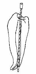
 The sugery involves removing the pulp tissue of a tooth which is either infected or in danger of infection due to an abcess at the apex of the tooth. The surgeon must drill into the crown, remove the pulp tissue from the canals, clean and shape each root, and seal the roots with a solid substance as near to the apex as possible. Failure to locate and clean every canal can result in failure, as can miscalculation at the filing stage, where the surgeon must carefully shape each canal without breaking through the dentine layer.
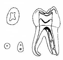
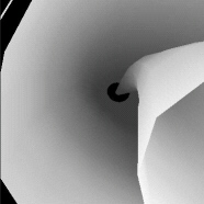
An additional difficulty involves the identification of the tooth's canal network. As shown in the figure on the right, a tooth can vary in the number, size and structure of the canals. Canals can also split apart, form a delta near the tooth's apex, gradually become more narrow and then wide again, etc. Some problems are sufficiently rare, while others occur more frequently, depending on the patient and the tooth in question.
We can use the benefits of computer graphics to provide an aid for dentists performing this surgery, and to give dental student additional instructional tools when learning the proper techniques. Ideally, a dentist would be able to have a 3D model of the given tooth's root network, as well as some numerical data of the width and depth of the canals. While modern X-rays do provide an overview of the root structure, they are insufficient for providing the detail necessary to give the dentist pertinent information. Instead, the surgeon must rely on experience and sensitivity to explore and shape the canals. Taking multiple X-rays would provide a more detailed level of information, but numerous X-rays in close proximity to the brain can be very harmful or fatal. Some other scanning technique is required to provide data which can be used to model the root network to a sufficient degree of accuracy.
For teaching, a student learning root canal surgery must rely on 2D images of tooth structures, and practise on extracted teeth or plastic models. What is lacking is twofold:
With only a limited number of practice models and extracted teeth available, the student may be ill-prepared for the kind of cases one is likely to face as the necessity for this type of surgery increases.
Graphics can be used for educational purposes by providing realistic 3-dimensional models of a tooth and its canal network which the student can view and interact with. In addition, a tool which can generate 3D tooth models with various alterations in the canal network can allow the student to examine and practise on several types of models and canal irregularities.
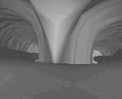
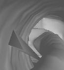
To create and display a 3D tooth model, the first step involves defining the method of data representation. Contours are one format which have been successful, particularly in other areas of medical imaging. Contour representation involves ordering a set of points into closed contours on parallel cross-sectional planes. The contours consist simply of vertices which combine to define an edge of the object. Contours must be closed, ordered, simple (non-self-intersecting) and must not intersect with other contours on the cross-sectional plane. Several contours can exist per cross-section, and contours can be embedded to one level, which represents a "hole" (or, in this case, the tooth's canal).
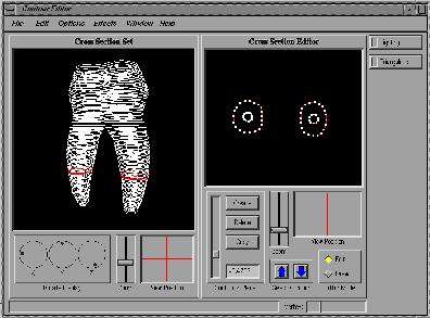 To create and modify contour data sets the Contour Editor was developed. The editor provides a view of the data set in 3D, as well as a view of one cross-section in 2D. The selected cross-section can be edited by selecting and moving a contour's vertex, and both contours and cross-sections can be copied, moved and deleted.
The two main features of the editor are contour interpolation and error checking:
Rendering the model from the contour data involves selecting an appropriate reconstruction method. The two main algorithms of Surface Reconstruction and Volume Rendering have both advantages and disadvantages:
This method involves generating a polygonal model from the given vertices for the surface. It is the more established technique given a data set of contours, and it renders the surface quickly. Accuracy can be a problem with some surface reconstruction algorithms, and traditional problems with correspondence, branching, tiling, and surface fitting.
This method has become more widely used as hardware technology increases to provide the computational power demanded by volume rendering algorithms. The process is best-suited for volume data, and produces a model which also renders the internal properties of the object as well as the surface. The old problems of speed are reduced with new powerful hardware systems, although the output of the object's internal density and substance is neither available with simple contour data, nor is it required when considering the data sets that are likely to be encountered with teeth.
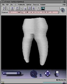

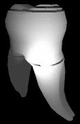
The reconstruction method chosen for this work was Nuages, a reconstruction method developed by Bernhard Geiger at the INRIA Research Institute in France. Nuages uses a combination of surface reconstruction and volume rendering principles to produce a surface rendering of the model in several output formats. The algorithm takes as input a contour data set, allowing for multiple contours per cross-section and embedded contours. The output is sufficiently good for testing and evaluation purposes, although even relatively simple data sets do produce the occasional error during reconstruction.
Nuages was set to output the rendered model in VRML. This modelling language provides the opportunity for users to view and interact with the object, and the additional portability and availability of VRML on the Internet gives this representation format increasing usefulness. If you have a VRML reader, you can walk through this Tooth VRML Model.
As a preliminary investigation into the use of computer graphics in endodontics, there is undoubtedly a high potential for a successful application of graphics to dentistry. Current techniques, while adequate, could be improved with newly available tools at both the learning level and the practical level. While the work outlined in this document merely samples the early stages of the possibilties, further work in this field has the potential to yield promising results.
Copyright © 2026 Mike Goetz